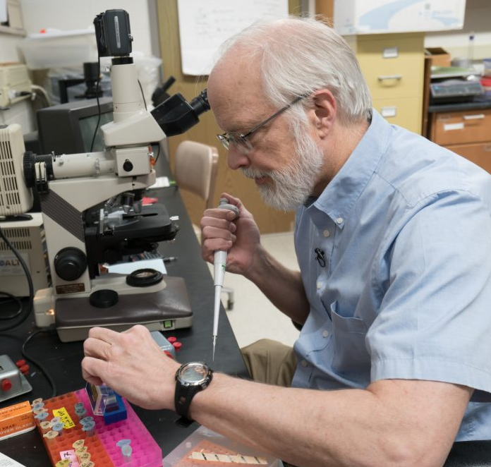
James Paulson, PhD

Description of the Research Program of Dr. J. Paulson
Dr. Paulson has retired, and is not working with any students during the pandemic.
Techniques and equipment used: Mammalian cell culture and cell cycle synchronization, light microscopy, isolation of chromosomes, electrophoresis and chromatography of proteins, enzyme assays.
Minimum experience: General chemistry, some biology or biochemistry.
Preferred experience: Organic chemistry, possibly quantitative analysis, some advanced biology or biochemistry lab work.
Current projects:
1) Specific induction of apoptosis by heat treatment of human cancer cells arrested in mitosis.
We have found that HeLa cells, a human cervical carcinoma cell line, will undergo apoptosis (physiological cell death) when arrested in metaphase of mitosis, treated with a compound that inhibits certain anti-apoptotic proteins (in the Bcl-2 family of proteins) and then shifted to higher temperature (42°C) -- called "hyperthermia". The occurence of apoptosis has been shown by morphological changes in the cells, suppression of those morphological changes by inhibitors of apoptosis, and by apoptosis-specific cleavage of certain proteins. Interphase cells are not affected by the same treatment.
Future work will focus on testing hypotheses concerning the possible mechanism behind this phenomenon. We are now trying to determine why treatment with the Bcl-2 inhibitor and hyperthermia induces apoptosis in metaphase-arrested cells but not in interphase cells. One clue comes from experiments suggesting that apoptosis only occurs when Cdk1-cyclin B (the enzyme that triggers mitosis and keeps cells in mitosis) is active.
2) Identification of protein phosphatases involved in exit from mitosis in yeast
As mentioned above, the enzyme Cdk1-cyclin B (also called MPF, for Mitosis Promoting Factor) has been shown to be responsible for initiating the events of mitosis. MPF causes many proteins to be modified by the addition of phosphate groups (this is called protein phosphorylation). This changes the function of those proteins, triggering the cellular changes that are characteristic of mitosis. Normally at the end of mitosis Cdk1-cyclin B (MPF) is inactivated by destruction of the cyclin subunit, and the proteins are dephosphorylated. We have shown, both in human cells and in yeast, that artificially inactivating or inhibiting Cdk1 in metaphase-arrested cells causes them to leave mitosis (without segregating their chromosomes or undergoing cytokinesis) and go back to interphase. During this transition, the proteins that were phosphorylated at the start of mitosis are dephosphorylated.
This observation provides a tool to study which protein phosphatase enzymes dephosphorylate proteins at the end of mitosis. Others have argued that the proteins phosphorylated by Cdk1 at mitosis are dephosphorylated by Cdc14 phosphatase. However, our experiments with HeLa cells and with budding yeast (Saccharomyces cerevisiae) point to Protein Phosphatase 1 (PPase 1, or Glc7 in yeast). Further work needs to be done to determine whether Cdc14 phosphatase is also involved.
3) Other studies on histone phosphorylation, mitotic chromosome structure, and chromosome isolation
We have made several other interesting observations over the past few years that merit further investigation. First, we found that treatment of cells with inhibitors of protein phosphatases (for example, inhibitors of PP1) induces nuclear envelope breakdown and "prematurely condensed chromosomes" (PCCs) – in other words, a sort of premature mitosis-like state. When this is done using G1-phase cells, the condensed chromosomes lack phosphorylated histone H1 (one of the major proteins in chromosomes). This shows that histone H1 phosphorylation is not required for chromosome condensation. We would like to use this system to test whether or not histone H3 phosphorylation is required for chromosome condensation.
Second, we have studied acetylation of the histones (the major chromosomal proteins), which means addition of acetyl groups to many of the lysine side chains of the proteins. If interphase HeLa cells are treated with inhibitors of histone deacetylases (HDACs), the histones become highly acetylated. However, they do not become highly acetylated when metaphase-arrested cells are treated in the same way. We are currently trying to determine the reasons for this. It could be that the enzymes that add the acetyl groups (histone acetyltransferases, or HATs) are down-regulated during mitosis, or it could be that the sites of acetylation are not accessible to the HATs in condensed mitotic chromosomes.
Third, we have found that inhibitors of the topoisomerase II do not prevent the formation of PCCs, but they do prevent the individualization of chromosomes. It remains to be worked out, using these inhibitors as tools, whether individualization of chromosomes occurs during chromosome condensation or later, when the chromosomes are separated at anaphase of mitosis.
Fourth, we have discovered some new "tricks" that might be useful for isolating mitotic chromosomes or preventing the dephosphorylation of histones and other proteins during the isolation. These need to be investigated further.
Collaborations:
(1) Dr. Linfeng Xie, University of Wisconsin Oshkosh; synthesizing organic compounds useful in studying chromosomes and mitosis.
(2) Dr. Bill Earnshaw and Dr. Kumiko Samejima, University of Edinburgh, Scotland; studies on mitotic chromosomes.

